නොදකින ලෝකයේ පින්තුර.. ( Nikon Small World Photomicrography Competition )
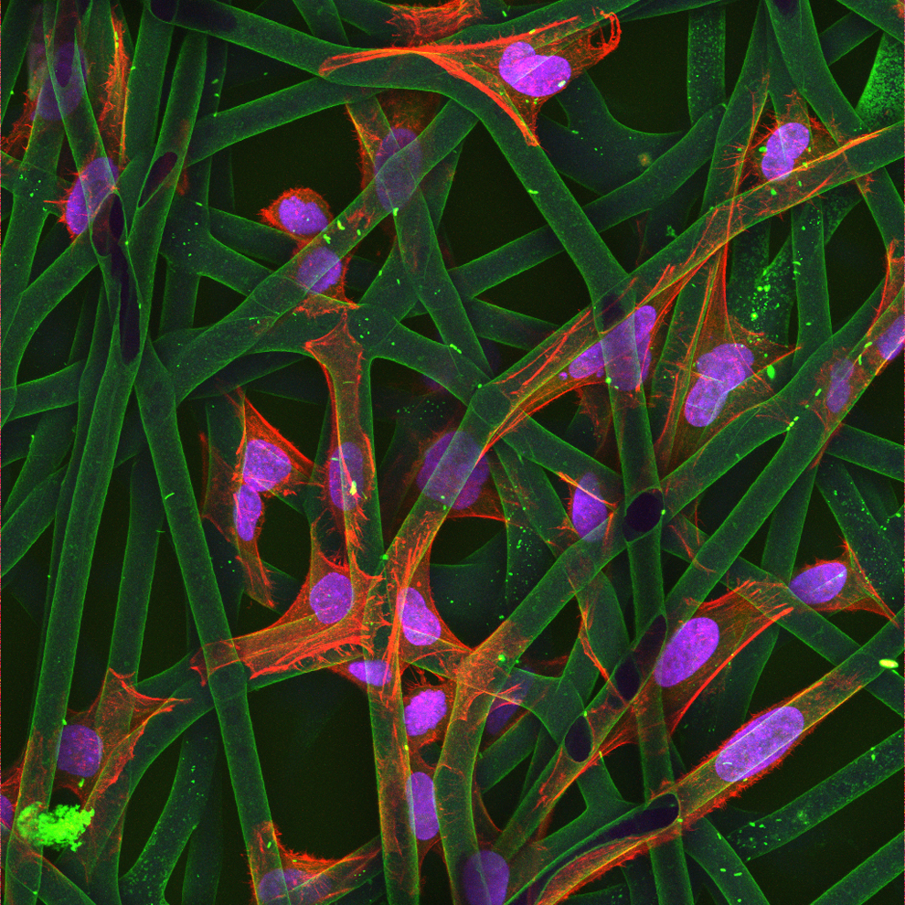
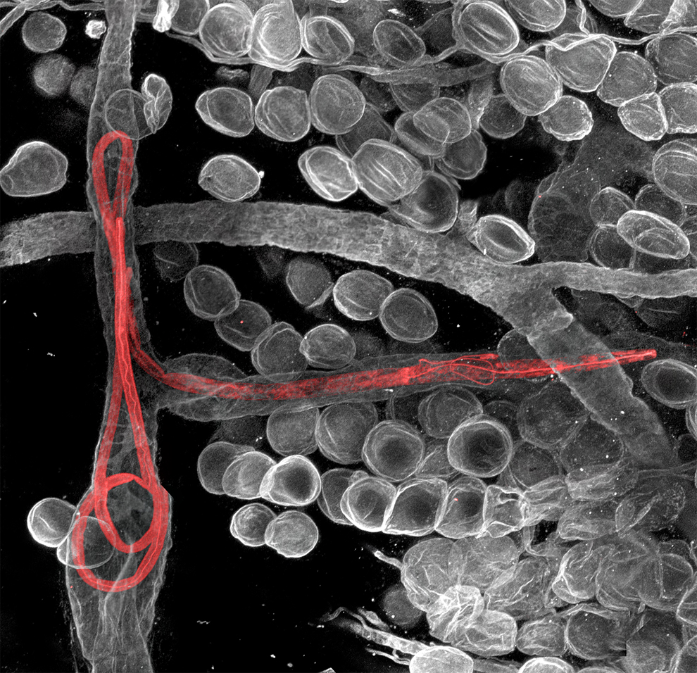
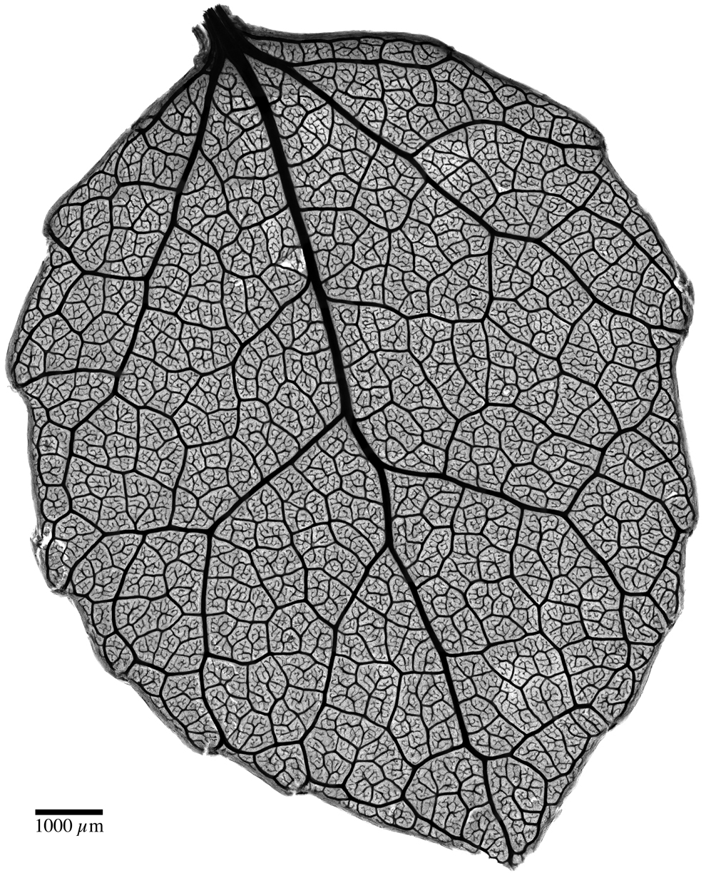
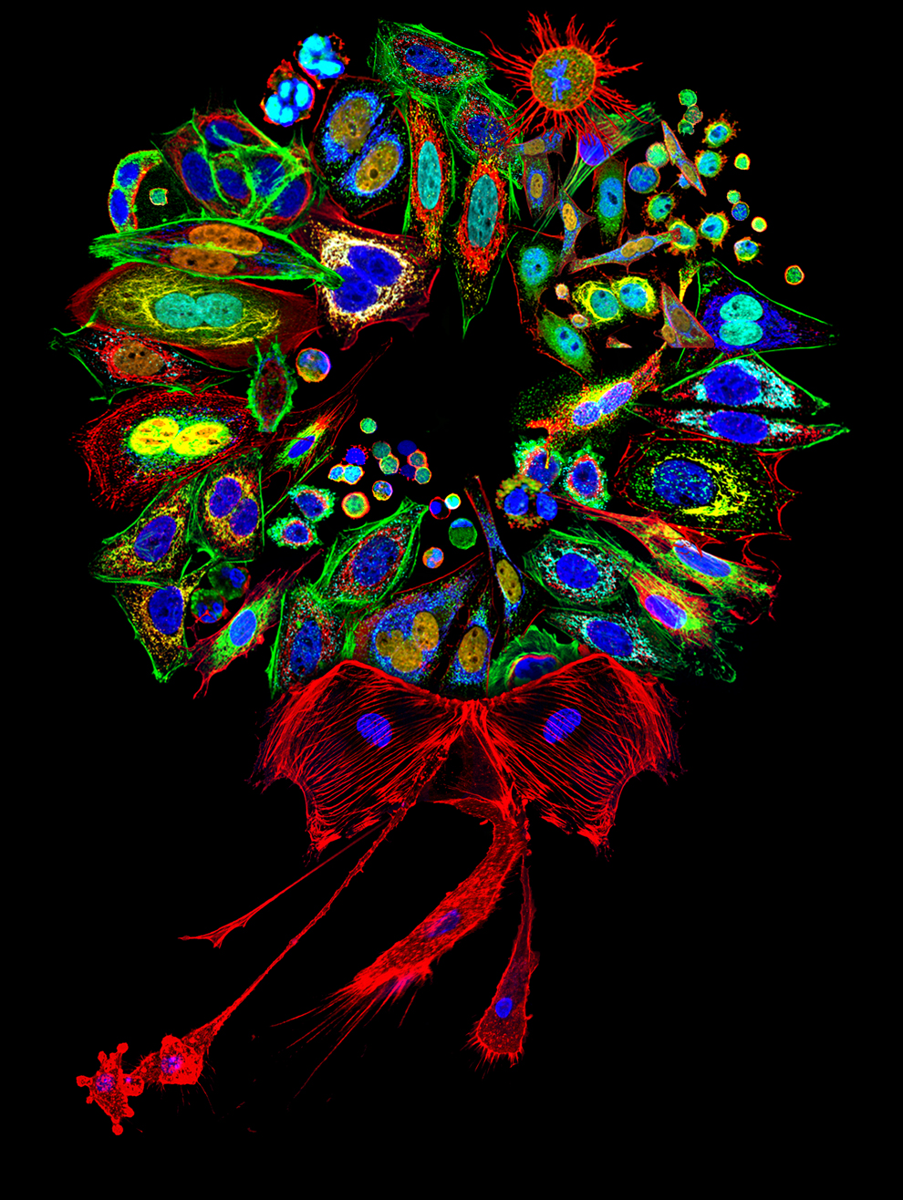
තරගයට 2011 දී ඉදිරිපත් කරලා හොදම පින්තුර විදිහට තේරුණ පින්තුර තමයි මේ..
පුදුමයි නේද ?? මේ හැම දේම අප අවට තියෙන දේවල්...
විශේෂත්වය තමයි ගොඩාක් සූම් කරලා..
ලොඩ් වෙන්න ටිකක් වෙලා යාවී..

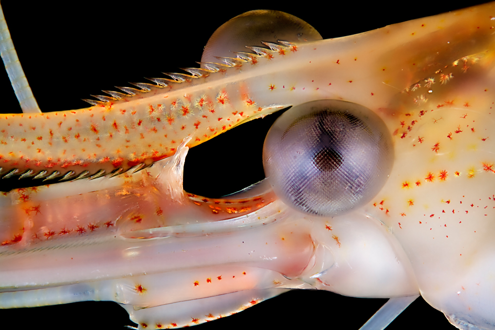
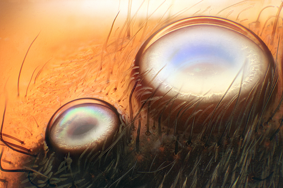
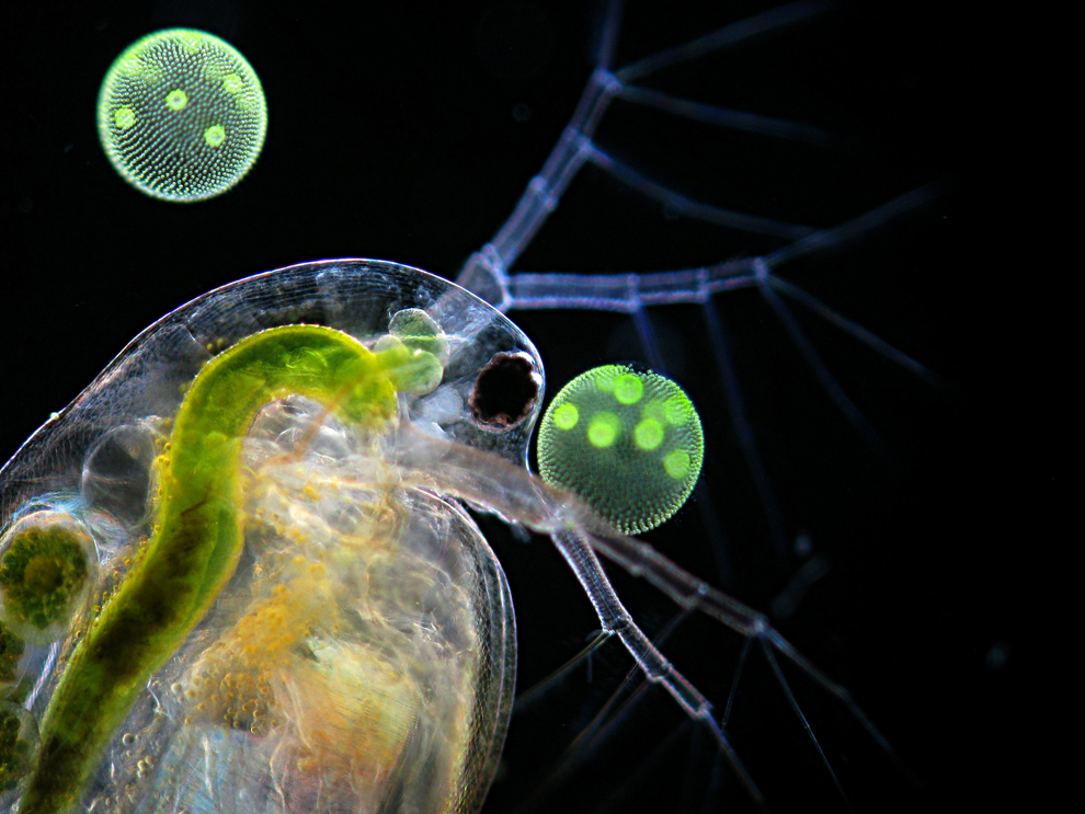
Wim van Egmond of the Micropolitan Museum in Rotterdam, Netherlands photographed a Leptodora kindtii (giant waterflea) eye from a living specimen using the differential interference contrast method. (Wim van Egmond)
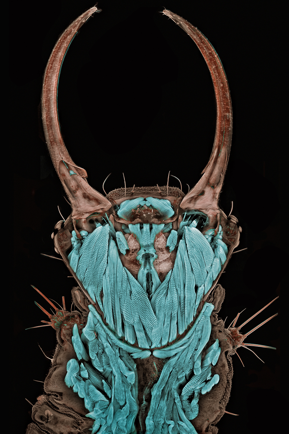
Dr. Igor Siwanowicz of the Max Planck Institute of Neurobiology in Martinsried, Germany shot "Portrait of a Chrysopa sp. (green lacewing) larva" at 20x magnification using the confocal method. (Dr. Igor Siwanowicz) #
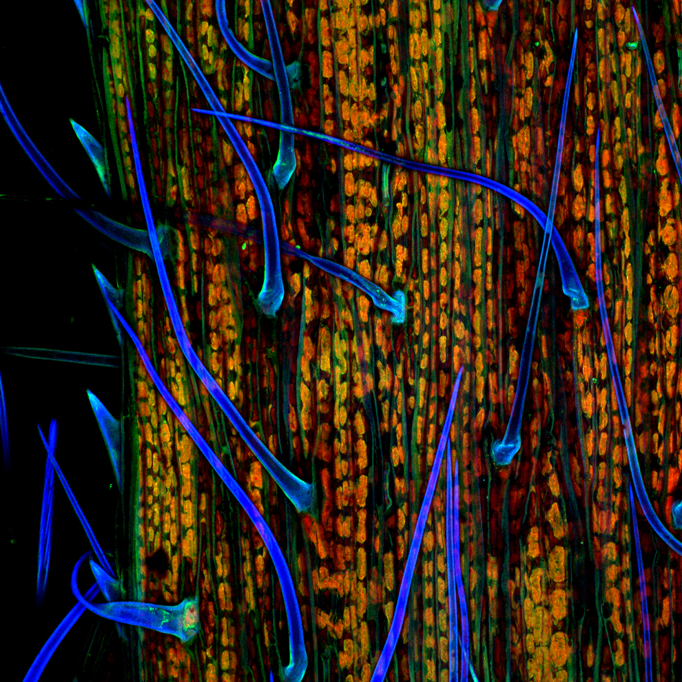
A blade of grass at 200x magnification by Dr. Donna Stolz of the University of Pittsburgh using confocal stack reconstruction and autofluorescence. (Dr. Donna Stolz) #
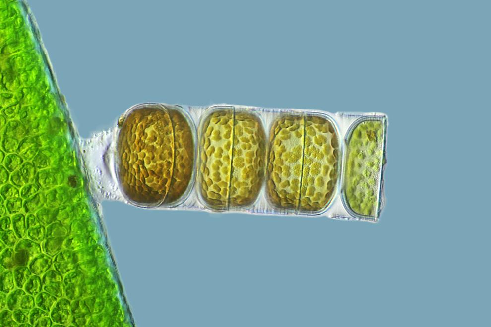
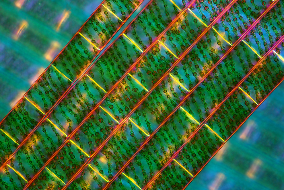
A living specimen of Melosira moniliformis at 320x magnification imaged with differential interference contrast by Frank Fox of Fachhochschule Trier in Trier, Rheinland-Pfalz, Germany. (Frank Fox) #
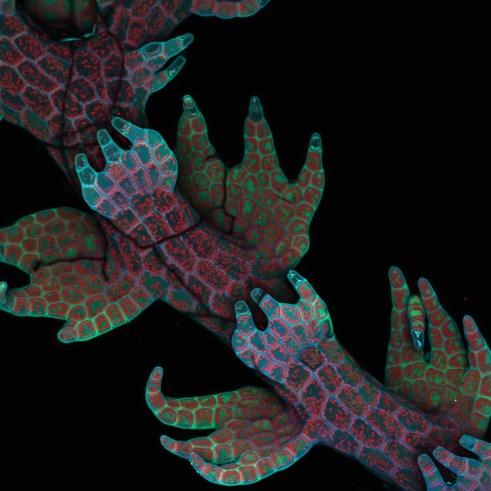
Intrinsic fluorescence in Lepidozia reptans (liverwort) at 20x magnification in a live mount with confocal microscopy by Dr. Robin Young of the University of British Columbia in Vancouver. (Dr. Robin Young) #
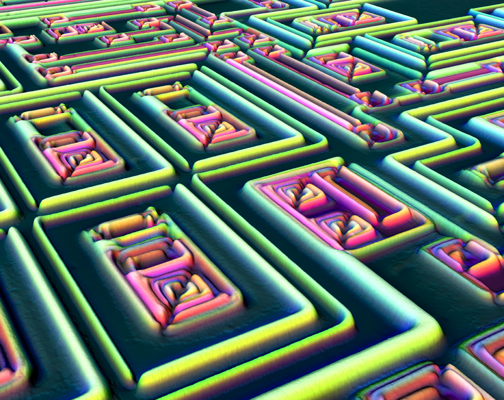
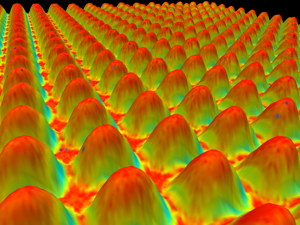
The surface of a microchip in a 3D reconstruction at 500x magnification using incident light and Normarski interference contrast by Alfred Pasieka of Germany. (Alfred Pasieka) #
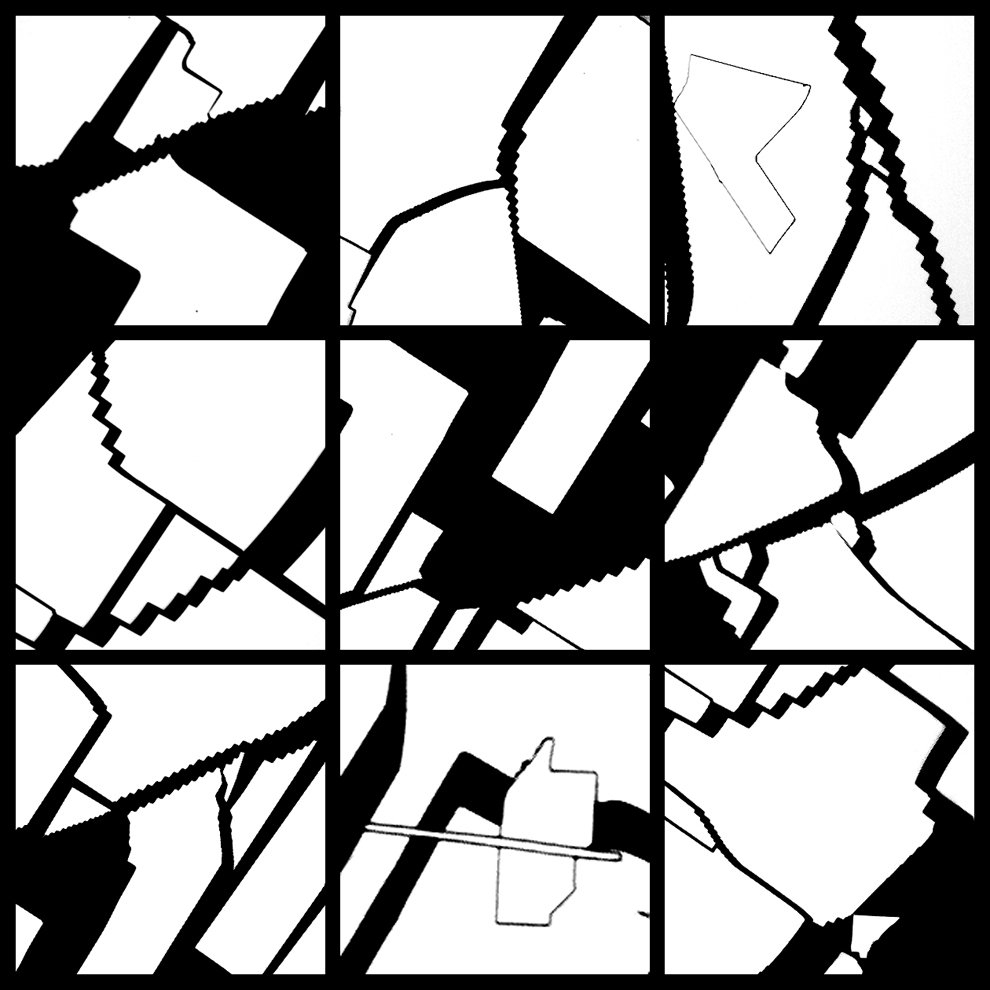
Cracked gallium arsenide solar cell films at 50x magnification using the brightfield method by Dennis Callahan of the California Institute of Technology in Pasadena. (Dennis Callahan) #
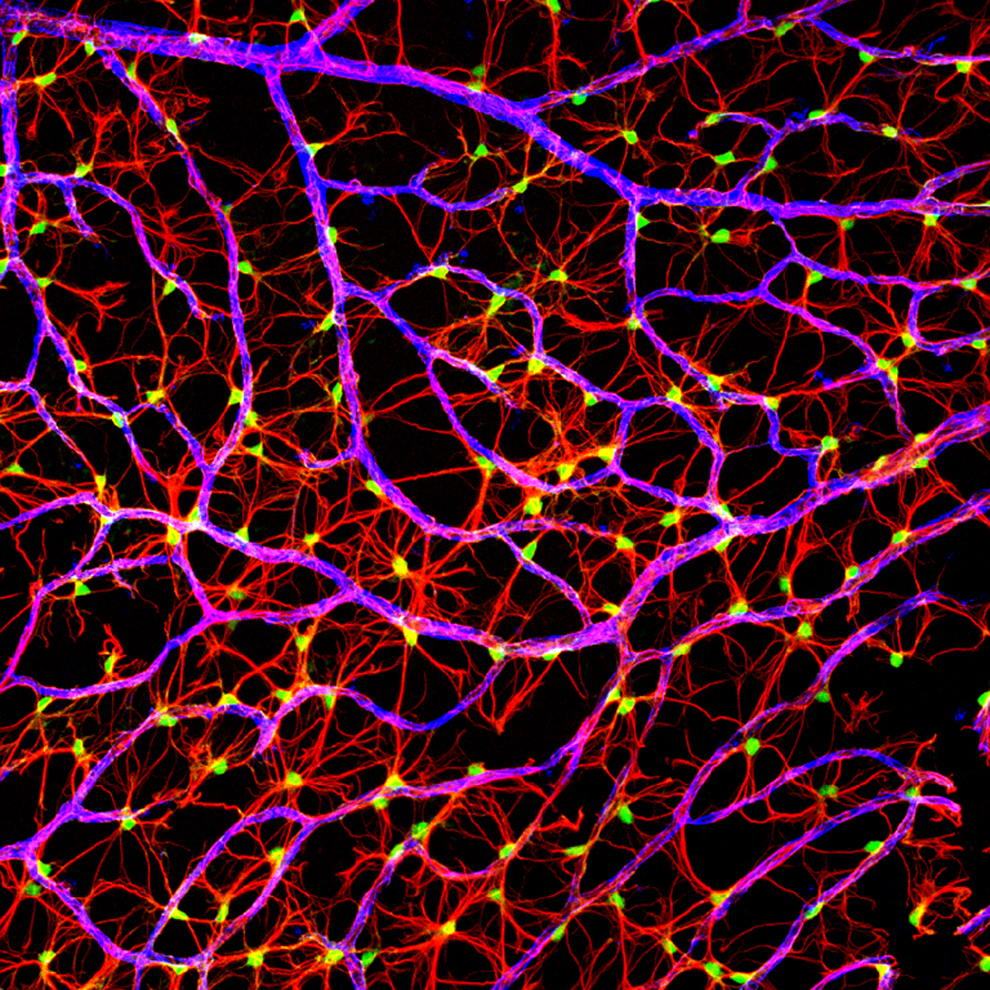
A mouse nerve fiber layer on a retinal flatmount at 40x magnification by Gabriel Luna of the UC Santa Barbara Neuroscience Research Institute in Santa Barbara, Calif. using the laser confocal scanning method. (Gabriel Luna) #
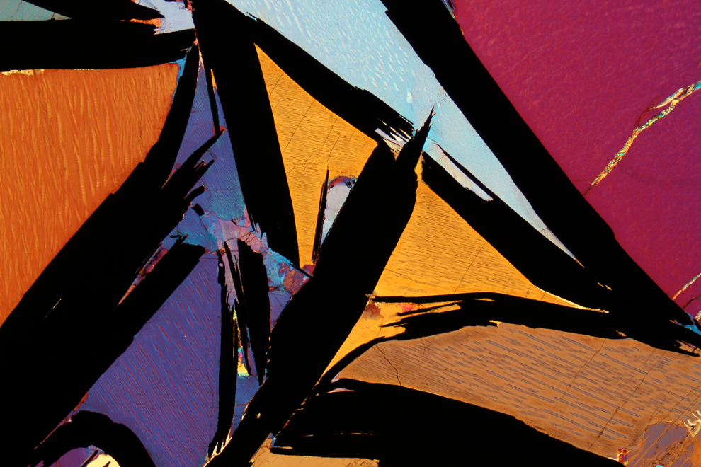
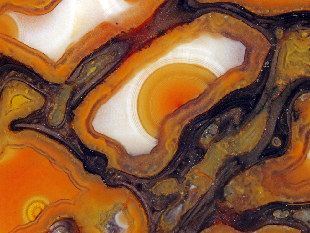
Graphite-bearing granulite from Kerala, India in polarized light at 2.5x magnification by Dr. Bernardo Cesare of the Department of Geosciences in Padova, Italy. (Dr. Bernardo Cesare) #
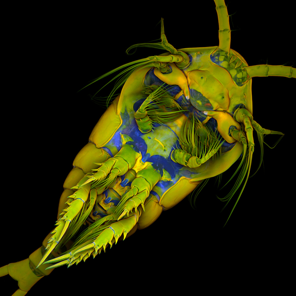
A marine copepod, Temora longicornis, at 10x magnification imaged by Dr. Jan Michels of Christian-Albrechts-Universität zu Kiel in Kiel, Germany using the confocal process with autofluorescence and Congo red fluorescence. (Dr. Jan Michels) #
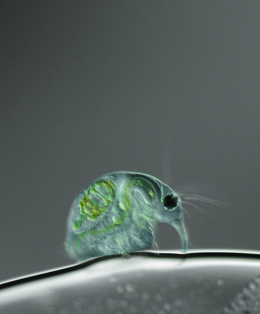
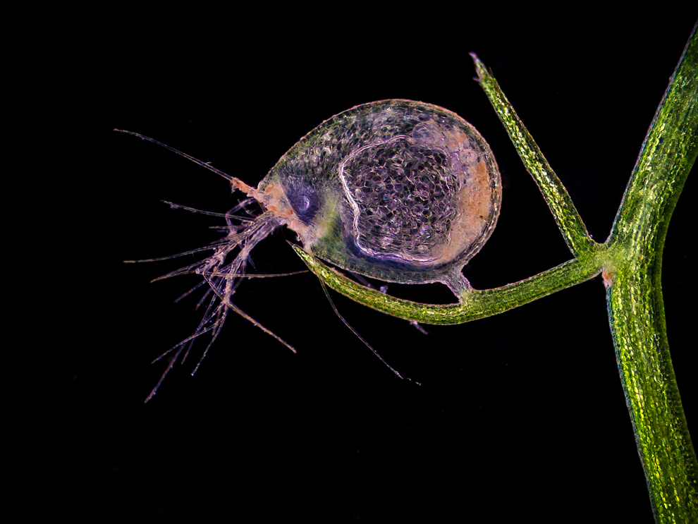
Joan Rohl of the Institute for Biochemistry and Biology in Potsdam, Germany used differential interference contrast to capture a freshwater water flea, Daphnia magna, at 100x magnification. (Joan Rohl) #
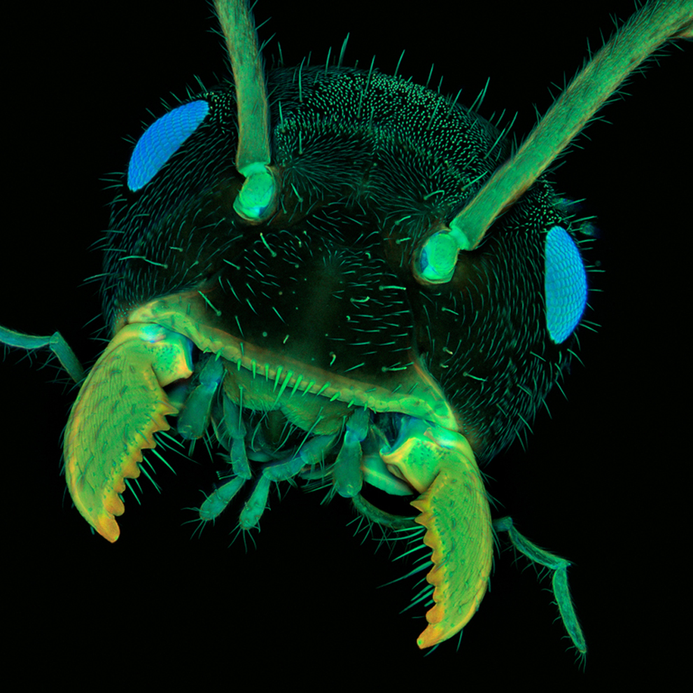
An ant head magnified ten times and photographed by Dr. Jan Michels of Christian-Albrechts-Universität zu Kiel in Kiel, Germany using the confocal method with autofluorescence. (Dr. Jan Michels) #
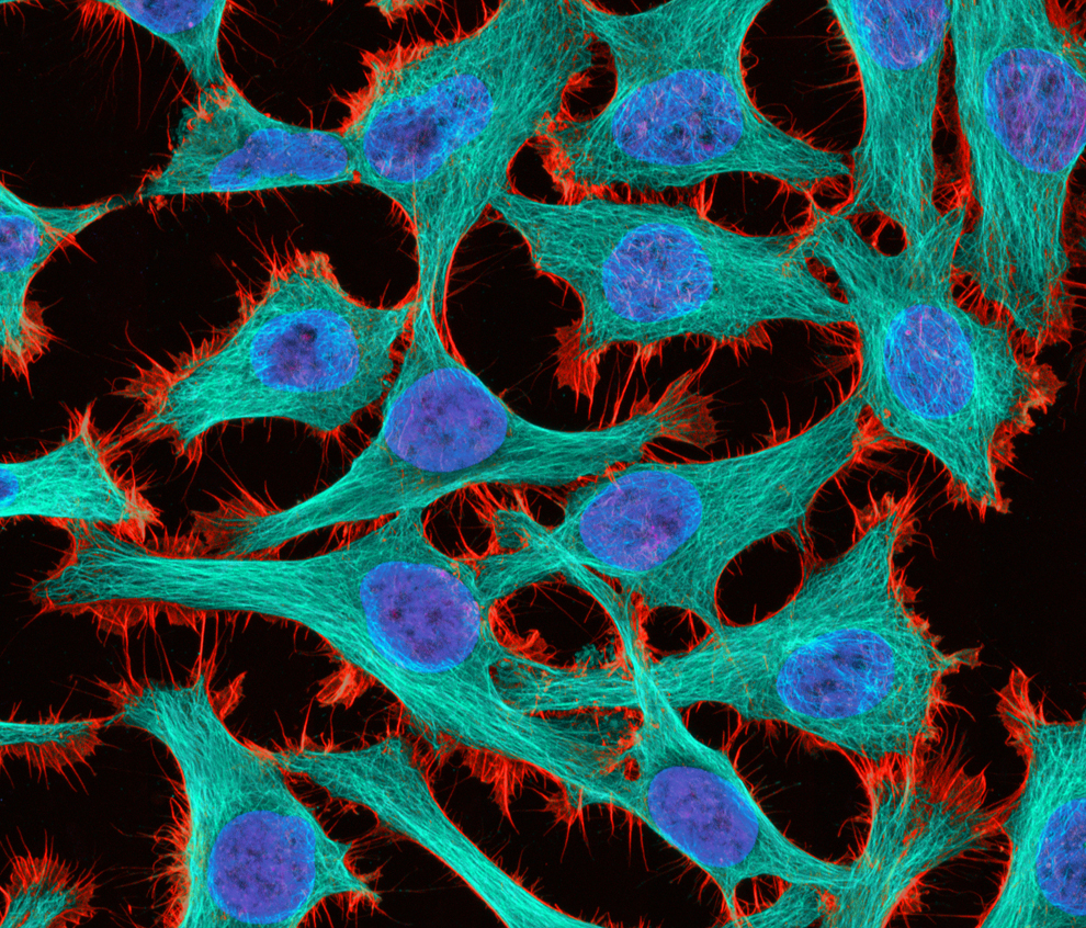
Thomas Deerinck of the National Center for Microscopy and Imaging Research in La Jolla, Calif. captured HeLa (cancer) cells at 300x magnification with 2-photon fluorescence. (Thomas Deerinck) #
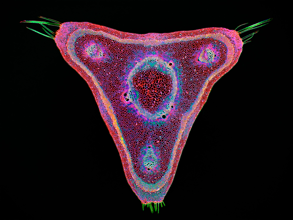
A curare vine (Chondrodendron tomentosum) in cross-section at 45x magnification was shot by Dr. Stephen S. Nagy of Montana Diatoms in Helena, Montana using the brightfield method and digitally inverted. (Dr. Stephen S. Nagy) #
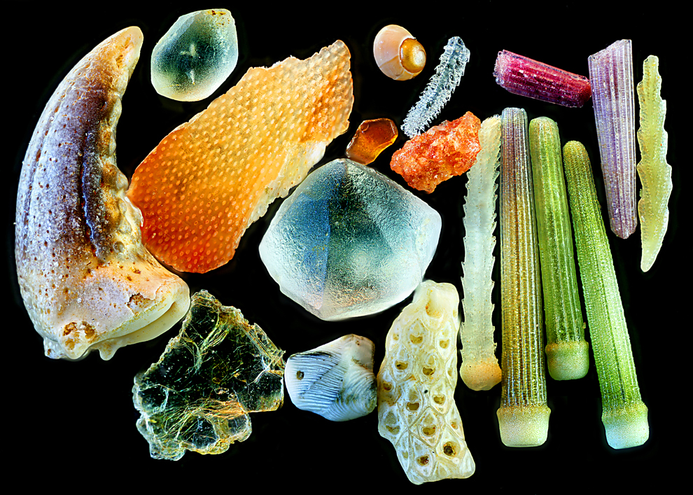
Using reflected light, Yanping Wang of the Beijing Planetarium in Beijing captured sand at 4x magnification. (Yanping Wang) #
Nikon Small World Photomicrography Competition
තරගයට 2011 දී ඉදිරිපත් කරලා හොදම පින්තුර විදිහට තේරුණ පින්තුර තමයි මේ..
පුදුමයි නේද ?? මේ හැම දේම අප අවට තියෙන දේවල්...
විශේෂත්වය තමයි ගොඩාක් සූම් කරලා..
ලොඩ් වෙන්න ටිකක් වෙලා යාවී..

Wim van Egmond of the Micropolitan Museum in Rotterdam, Netherlands photographed a Leptodora kindtii (giant waterflea) eye from a living specimen using the differential interference contrast method. (Wim van Egmond)
Dr. Igor Siwanowicz of the Max Planck Institute of Neurobiology in Martinsried, Germany shot "Portrait of a Chrysopa sp. (green lacewing) larva" at 20x magnification using the confocal method. (Dr. Igor Siwanowicz) #
A blade of grass at 200x magnification by Dr. Donna Stolz of the University of Pittsburgh using confocal stack reconstruction and autofluorescence. (Dr. Donna Stolz) #
A living specimen of Melosira moniliformis at 320x magnification imaged with differential interference contrast by Frank Fox of Fachhochschule Trier in Trier, Rheinland-Pfalz, Germany. (Frank Fox) #
Intrinsic fluorescence in Lepidozia reptans (liverwort) at 20x magnification in a live mount with confocal microscopy by Dr. Robin Young of the University of British Columbia in Vancouver. (Dr. Robin Young) #
The surface of a microchip in a 3D reconstruction at 500x magnification using incident light and Normarski interference contrast by Alfred Pasieka of Germany. (Alfred Pasieka) #
Cracked gallium arsenide solar cell films at 50x magnification using the brightfield method by Dennis Callahan of the California Institute of Technology in Pasadena. (Dennis Callahan) #
A mouse nerve fiber layer on a retinal flatmount at 40x magnification by Gabriel Luna of the UC Santa Barbara Neuroscience Research Institute in Santa Barbara, Calif. using the laser confocal scanning method. (Gabriel Luna) #
Graphite-bearing granulite from Kerala, India in polarized light at 2.5x magnification by Dr. Bernardo Cesare of the Department of Geosciences in Padova, Italy. (Dr. Bernardo Cesare) #
A marine copepod, Temora longicornis, at 10x magnification imaged by Dr. Jan Michels of Christian-Albrechts-Universität zu Kiel in Kiel, Germany using the confocal process with autofluorescence and Congo red fluorescence. (Dr. Jan Michels) #
Joan Rohl of the Institute for Biochemistry and Biology in Potsdam, Germany used differential interference contrast to capture a freshwater water flea, Daphnia magna, at 100x magnification. (Joan Rohl) #
An ant head magnified ten times and photographed by Dr. Jan Michels of Christian-Albrechts-Universität zu Kiel in Kiel, Germany using the confocal method with autofluorescence. (Dr. Jan Michels) #
Thomas Deerinck of the National Center for Microscopy and Imaging Research in La Jolla, Calif. captured HeLa (cancer) cells at 300x magnification with 2-photon fluorescence. (Thomas Deerinck) #
A curare vine (Chondrodendron tomentosum) in cross-section at 45x magnification was shot by Dr. Stephen S. Nagy of Montana Diatoms in Helena, Montana using the brightfield method and digitally inverted. (Dr. Stephen S. Nagy) #
Using reflected light, Yanping Wang of the Beijing Planetarium in Beijing captured sand at 4x magnification. (Yanping Wang) #